
7. Biceps Femoris
Origin:
- Long head: Ischial tuberosity
- Short head: Linea aspera and lateral supracondylar line of the femur
Insertion: Lateral aspect of the fibular head (split by the lateral collateral ligament)
Action:
- Flexion of the knee
- Lateral rotation of the knee
- Extension of the hip on a stable lower leg
Innervation:
- Long head: Tibial division of the sciatic nerve (L5/S1/S2)
- Short head: Peroneal division of the sciatic nerve (L5/S1/S2)

Needling
Position: Prone
Palpation: Palpate the biceps femoris lateral to midline on the posterior aspect of the femur – The short head can be palpated up from its insertion point to the lateral supracondylar ridge of the femur
Technique:
-
- Needle perpendicular to the femur lateral to midline
- Needle in a slight oblique direction medial to lateral avoiding the midline of the posterior thigh
- To needle the short head of biceps femoris, needle more distal along the lateral supracondylar ridge of the femur – Needle perpendicularly using the lateral aspect of the femur as a backdrop
Caution: Low – the sciatic nerve lies directly in the midline of the posterior thigh
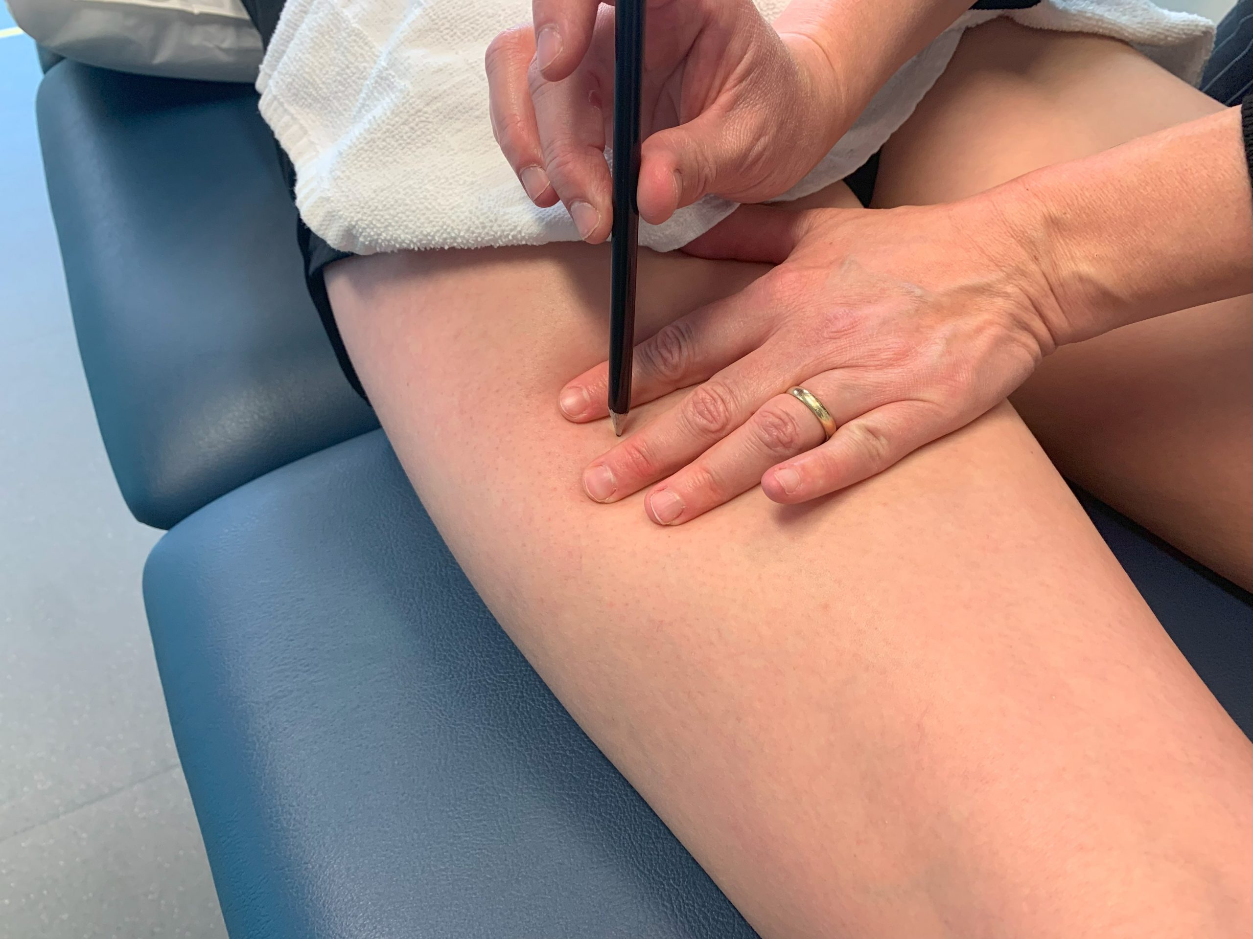
Video 1.8 Needling biceps femoris, original video by Nadine Crocker, CC BY-NC 4.0.
Video 1.9 Needling biceps femoris (short head), original video by Nadine Crocker, CC BY-NC 4.0.