
Overview: Stomach Sinew Channel (Leg Yangming)
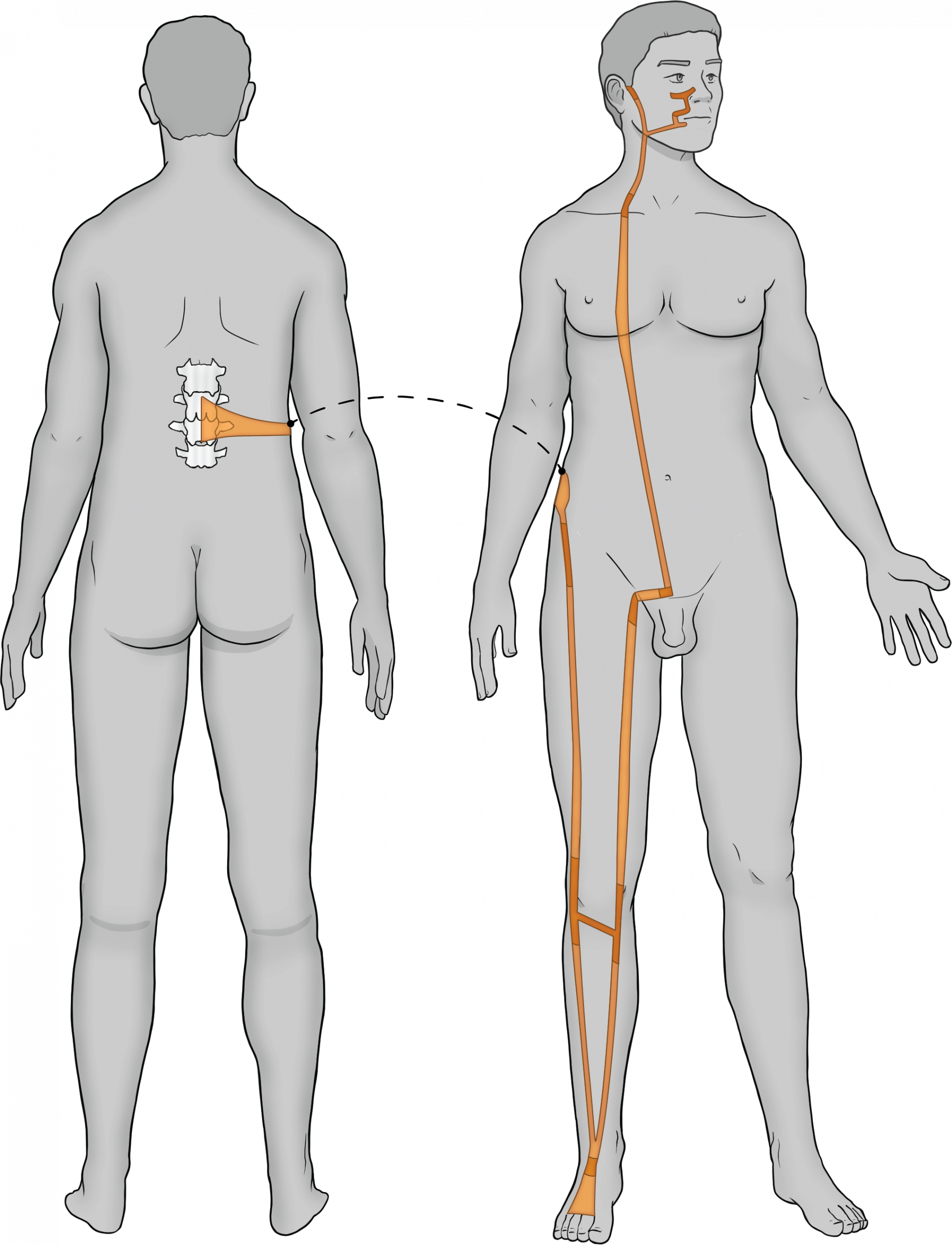
The Stomach Sinew Channel: Trajectory and associated anatomy
-
- The Stomach sinew channel begins on the dorsum of the toes and travels up the foot and continues up the anterior compartment of the lower leg to bind just inferior to the knee on the anterolateral aspect. There is also another divergent beginning at the insertion of the tibialis anterior on the medial aspect of the foot, which will blend with the anterior compartment of the lower leg as well. (Anatomy: Dorsal interossei, Extensor digitorum brevis, Tibialis anterior, Extensor hallucis longus, Extensor digitorum longus, Peroneus tertius)
-
- One branch will bind at the anterior inferior iliac spine (AIIS) via most of the quadriceps.
- The other branch will bind at the anterior superior iliac spine (ASIS) via the rectus femoris and the sartorius. From here, this channel will branch. Both branches will take a similar trajectory through the patella tendon to the anterior thigh. (Anatomy: A) Vastus lateralis, Vastus medialis, Vastus intermedius B) Rectus femoris, Sartorius)
- The stomach Sinew Channel will then travel to the pubis via the inguinal ligament and then up the abdomen via the rectus abdominis. All the lateral abdominal muscles also bind to the inguinal ligament and travel up to bind to the lower rib cage. (Anatomy: Inguinal ligament, Rectus abdominis, External oblique, Internal oblique, Transverse abdominis)
- The stomach Sinew Channel will continue up the midline of the body via the sternalis (if present) and continue up the neck via the voluntary muscles of swallowing. It continues through the voluntary muscles of the soft (lower) palate binding on the mandible (including the tongue). (Anatomy: Sternalis, Omnohyoid, Sternothyroid, Sternohyoid, Digastric, Mylohyoid)
- From inside the mandible, the channel emerges to join the muscles of the chin and lower lip, continuing around the mouth to attach to the cheek and nose including the muscles of the lower eyelid. (Anatomy: Mentalis, Depressor labii inferioris, Depressor anguli oris, Levator anguli oris, Orbicularis oris, Levator labii superioris alaeque nasi, Risorius, Zygomaticus minor and major, Orbiuclaris oculi)