
2. Extensor Digitorum Brevis
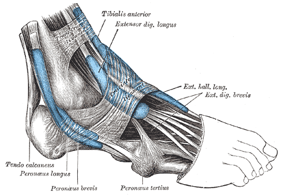
Origin: Superior lateral aspect of the calcaneus, the floor of the tarsal sinus, the talocalcaneal ligament and the inferior extensor retinaculum
Insertion: Base of digit 1, middle phalanx of digit 2-4 via the tendons of extensor digitorum longus
Action: Assists in extension of digit 1-5
Innervation: Deep fibular nerve (S1/2)
Needling
Position: Prone
Palpation: Palpate the lateral aspect of the dorsum of the foot – Resist toe extension to help isolate this muscle
Technique: Needle directly perpendicular to this muscle on the dorsum of the foot
Caution: Low
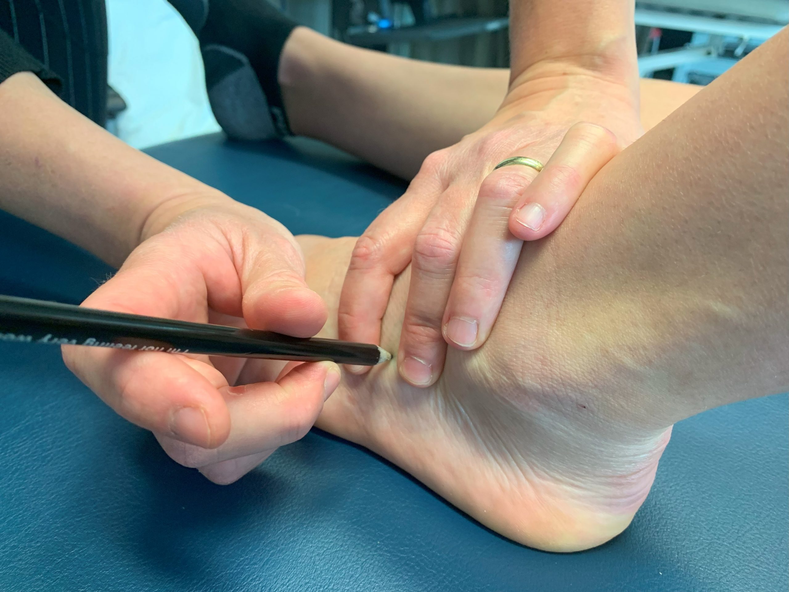
Video 3.2 Needling of extensor digitorum brevis, original video by Nadine Crocker, CC BY-NC 4.0.