3. Tibialis Anterior

Origin: Lateral Tibial condyle, superior half of the lateral tibial surface and interosseous membrane

Insertion: Dorsal base of the first metatarsal and medial aspect of medial cuneiform

Action:

  • Dorsiflexion of ankle
  • Supination of foot

Innervation: Deep peroneal nerve (L4/5)
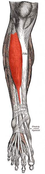
Anterior leg muscles (tibialis anterior).
Figure 3.6 Anterior leg muscles (tibialis anterior), adapted by Uwe Gille from the original image by Henry Gray and Henry Vandyke Carter (Public domain).
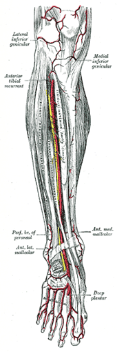
Arteries of the lower leg, anterior view.
Figure 3.7 Neurovascular bundle (tibialis anterior artery and vein, deep peroneal nerve), by Henry Gray and Henry Vandyke Carter (Public domain).

Needling

Position: Supine

Palpation: Palpate this muscle on the anterior aspect of the lower leg, resist supination to help isolate this muscle

Technique: Needle perpendicular to the tibialis anterior

Caution: Low – deep peroneal nerve lies just deep to this muscle
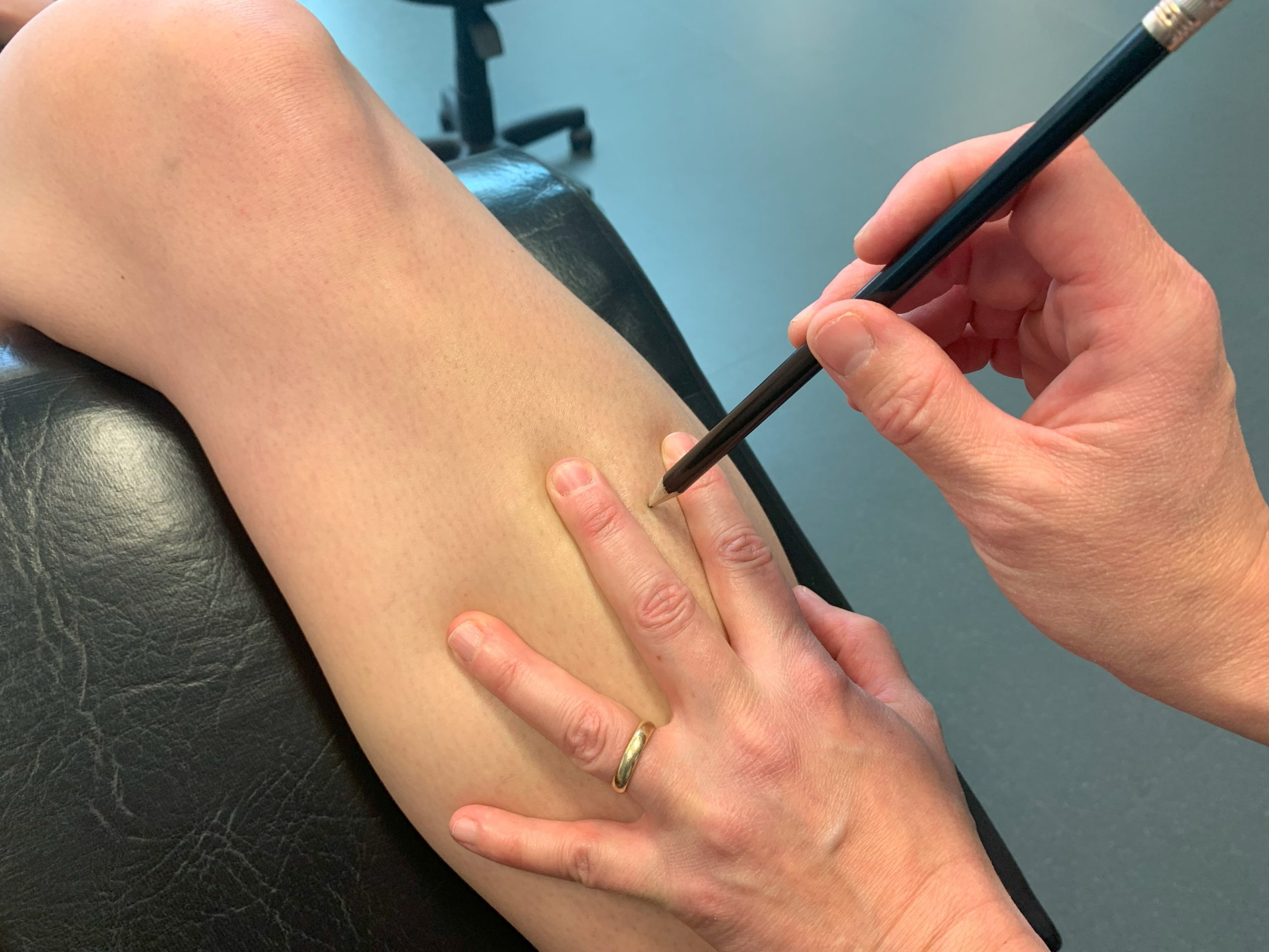
Needling tibialis anterior.
Figure 3.8 Needling tibialis anterior, original image by Nadine Crocker, CC BY-NC 4.0.

Video 3.3 Needling tibialis anterior, original video by Nadine Crocker, CC BY-NC 4.0.

License

Icon for the Creative Commons Attribution-NonCommercial 4.0 International License

Sinew Channel Needling Copyright © 2026 by Nadine Crocker is licensed under a Creative Commons Attribution-NonCommercial 4.0 International License, except where otherwise noted.