
11. Sartorius
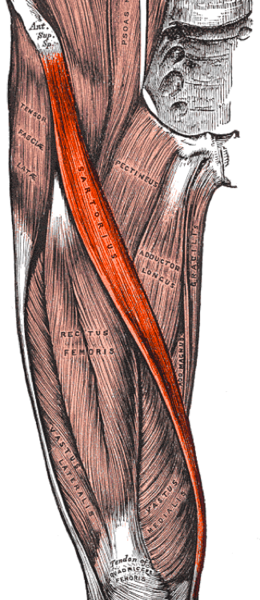
Origin: Anterior superior iliac spine
Insertion: Medial superior surface of the tibia (part of the pes anserine group)
Action:
- Flexes the hip
- Abducts the hip
- Lateral rotation of the hip
- Flexes the knee
Innervation: Femoral nerve (L2/3)
Needling
Position: Supine
Palpation: Resist hip flexion, abduction, and lateral rotation as well as knee flexion to isolate this muscle – Palpate along the front of the thigh in a diagonal direction from the anterior superior iliac spine to the medial aspect of the knee
Technique: Needle perpendicular to this muscle using the femur as a bony backdrop
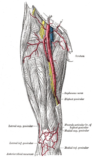
Caution: Moderate – this muscle crosses the femoral artery as it enters the adductor cannel (specifically the middle 1/3 of this muscle)
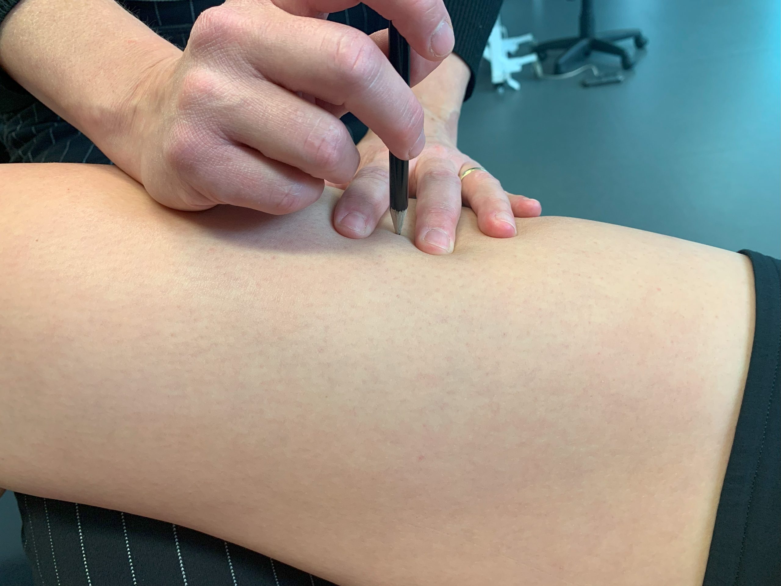
Video 3.11 Needling sartorius, original video by Nadine Crocker, CC BY-NC 4.0.