5. Lateral Pterygoid

Origin:

  1. Superior head: Infratemporal surface of sphenoid bone
  2. Inferior head: Lateral pterygoid plate (temporal bone)

Insertion: Neck of the mandible, articular disc, and capsule of the temporomandibular joint

Action:

  1. Bilaterally, they protrude the mandible and depress the chin
  2. Unilaterally, they produce lateral deviation of the jaw

Innervation: Lateral pterygoid nerve via the mandibular nerve via the trigeminal nerve (CN V)
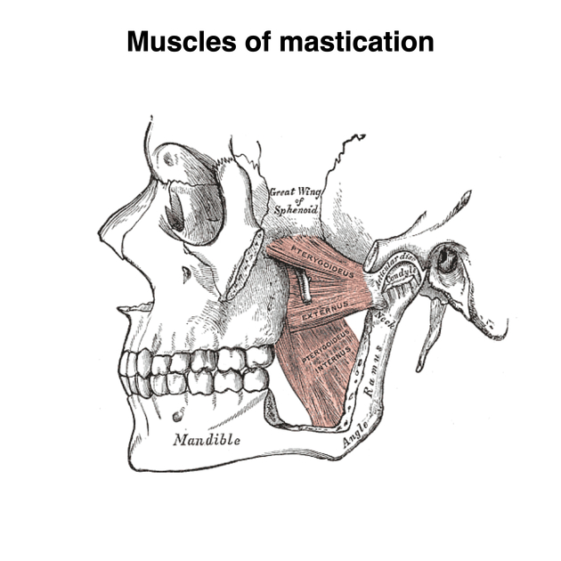
Lateral pterygoid.
Figure 8.11 Lateral pterygoid, by Henry Gray and Henry Vandyke Carter (Public domain).
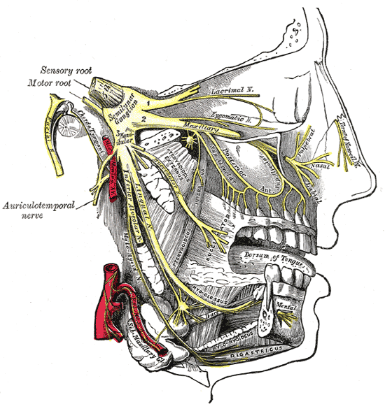
Distribution of the maxillary and mandibular nerves, and the submaxillary ganglion.
Figure 8.12  Trigeminal nerve, by Henry Gray and Henry Vandyke Carter (Public domain).
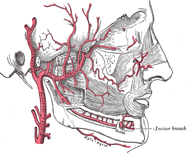
Blood supply.
Figure 8.13 Blood supply, by Henry Gray and Henry Vandyke Carter (Public domain).

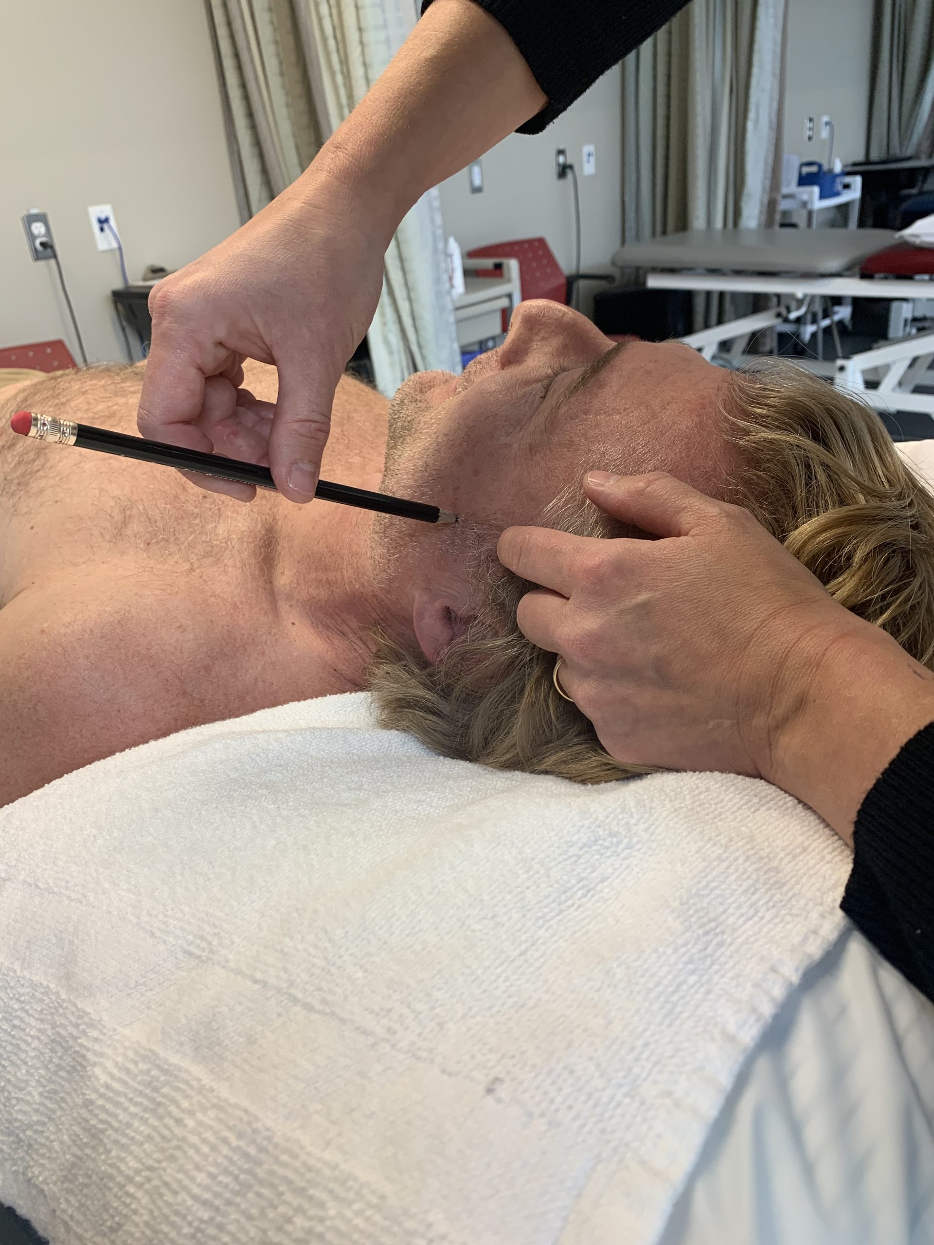
Needling

Position: Supine (often with the head turned to the side)

Palpation: Palpate the zygomatic arch and the hollow just inferior to it and anterior to the tragus (Stomach 7)

Technique: Directly perpendicular

Caution: Moderate
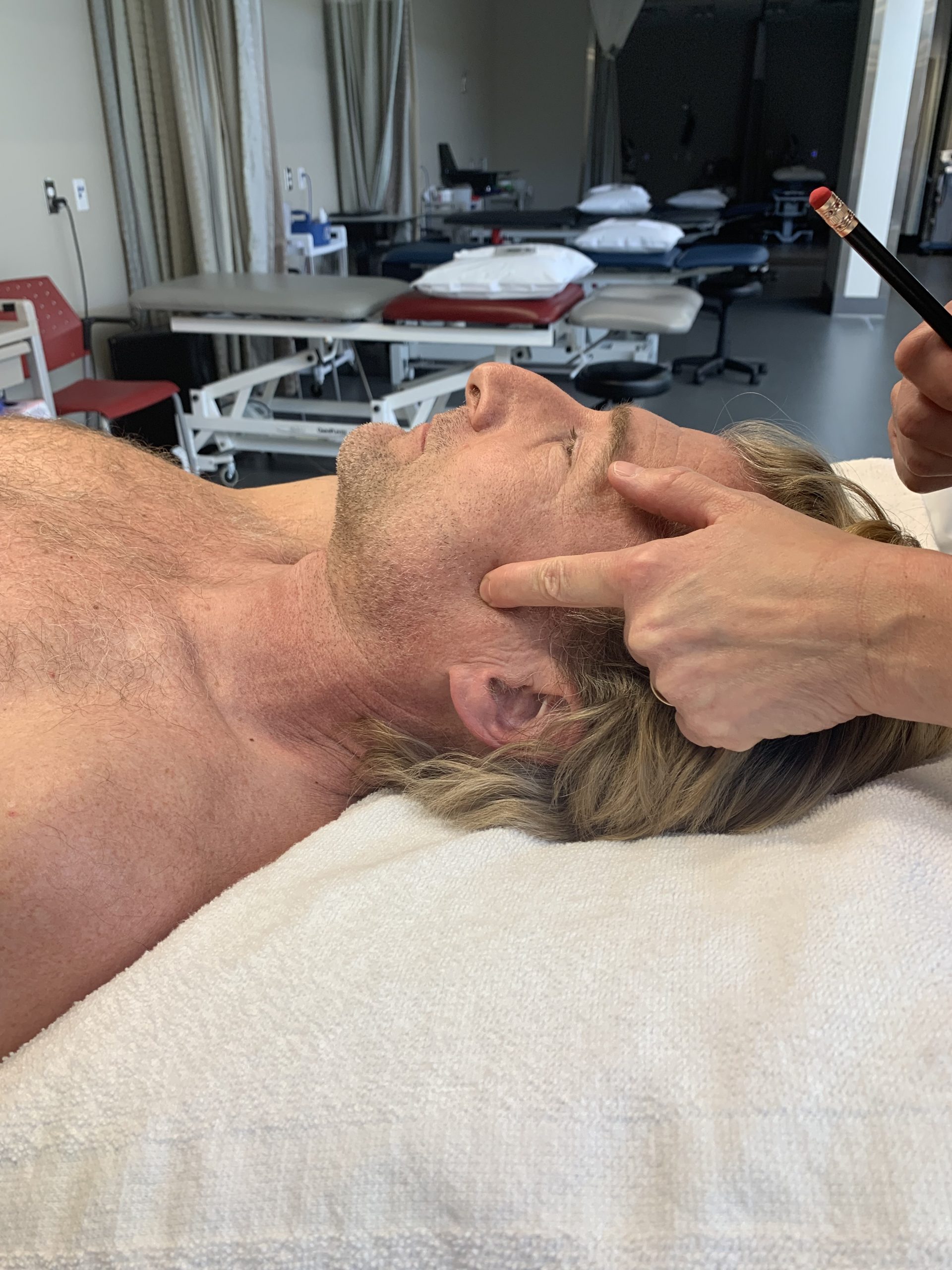
Needling lateral pterygoid.
Figure 8.14 Needling lateral pterygoid, original image by Nadine Crocker, CC BY-NC 4.0.
Needling lateral pterygoid.
Figure 8.15 Needling lateral pterygoid, original image by Nadine Crocker, CC BY-NC 4.0.

Video 8.6 Needling lateral pterygoid, original video by Nadine Crocker, CC BY-NC 4.0.

License

Icon for the Creative Commons Attribution-NonCommercial 4.0 International License

Sinew Channel Needling Copyright © 2026 by Nadine Crocker is licensed under a Creative Commons Attribution-NonCommercial 4.0 International License, except where otherwise noted.