
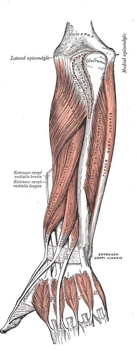
3. Extensor Pollicis Longus
Origin: Posterior surface of the ulna, radius, and interosseous membrane
Insertion: Base of distal phalanx of the thumb
Action: Extends the distal phalanx of the thumb at the metacarpophalangeal joint and interphalangeal joint
Innervation: Posterior interosseous nerve (C7/C8)
Needling
Position: Various (sitting, supine, prone) but with arm pronated
Palpation: Palpate approximately the middle 1/3 of the dorsal aspect of the forearm – Isolate muscle by asking patient to extend thumb
Technique: Needle perpendicular between radius and ulna
Caution: Low
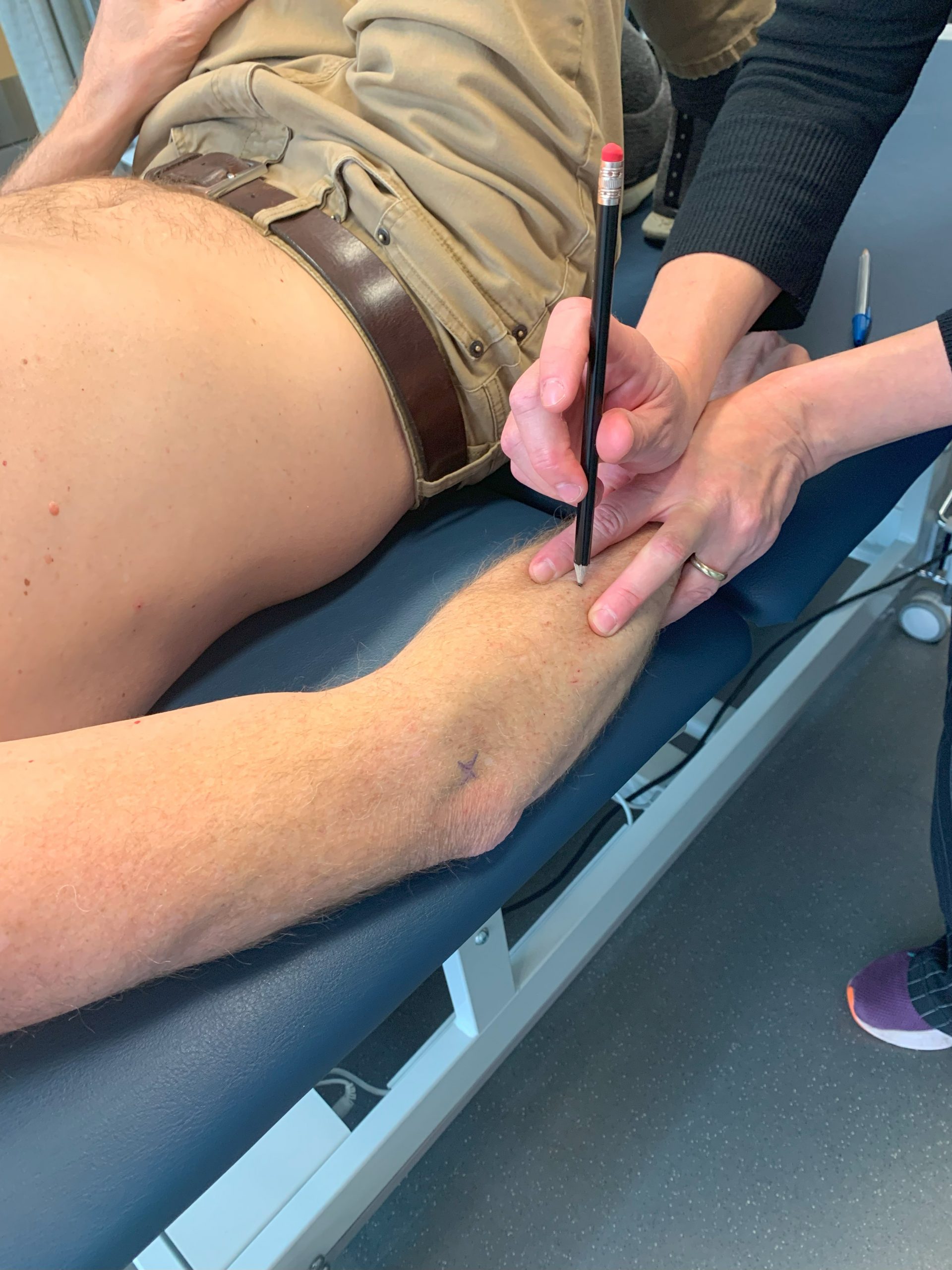
Video 9.3 Needling extensor pollicis longus, original video by Nadine Crocker, CC BY-NC 4.0.